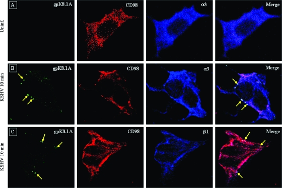
FIG. 6.
Confocal microscopic analysis of the interaction of KSHV with CD98 and α3/β1 integrin in infected cells. HMVEC-d were infected with KSHV at an MOI of 10 at 37°C for 10 min. The cells were fixed in 4% paraformaldehyde, permeabilized, and blocked. Triple-color confocal microscopy was performed on the uninfected (Uninf.) (A) and infected cells using anti-gpK 8.1A antibody for the detection of virus and anti-CD98 and anti-α3 (B) or anti-β1 (C) antibody for the detection of receptors. gpK8.1A was visualized with secondary antibodies conjugated to Alexa 488 (green), CD98 was visualized with Alexa 594 (red), and α3 or β1 integrin was visualized with Alexa 647 (blue). The overlays show the association of KSHV with CD98 and α3 or β1 integrin. The arrows indicate virus and the site of association of virus with CD98 and α3 or β1 integrin. Magnification, ×80.

