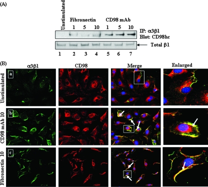
FIG. 7.
Effects of fibronectin and CD98 MAb on α3β1-CD98 association in HMVEC-d. (A) HMVEC-d were left unstimulated (top, lane 1), stimulated with fibronectin (10 μg/ml) for different time periods (minutes) (top, lanes 2 to 4), or stimulated with anti-CD98 MAbs (10 μg/ml) for different time periods (top, lanes 5 to 7). The cells were lysed, and the lysates were immunoprecipitated by anti-α3β1 integrin antibody and then blotted with CD98hc antibody. Loading was verified by blotting with total β1 integrin (bottom). (B) Effects of fibronectin and CD98 MAb on an α3β1-CD98 colocalization immunofluorescence assay in HMVEC-d. Cells unstimulated (a) or stimulated with CD98 MAb (10 μg/ml) (b) or with fibronectin (10 μg/ml) (c) were incubated with antibodies against α3β1 and CD98, and the staining was observed by incubation with Alexa 488 (green) for α3β1 and Alexa 594 (red) for CD98. The arrows indicate colocalization of activated α3β1 with CD98. Magnification, ×40. The boxed areas are enlarged in the rightmost column.

