FIG. 1.
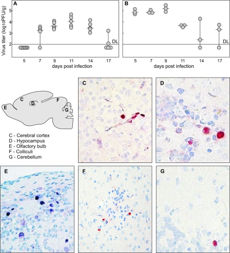
Kinetics of virus replication and spread in brain of MCMV-infected newborn mice. MCMV replication in the brains (A) and livers (B) of newborn mice. Mice were inoculated i.p. with 500 PFU of MCMV, 6 to 12 h after birth, and viral titers in their organs were assessed at various time points p.i. Productive infection in the brains was detected starting from day 7 p.i. and was visible through day 17 p.i. Titers of virus in individual mice (circles) and median values (horizontal bars) are shown. DL, detection limit. (C to G) MCMV antigen expression in the brains of newborn mice. Scattered infected cells located in the cerebral cortex, hippocampus, olfactory bulb, colliculi, and cerebellar cortex of infected mice are shown, as indicated on the figure, at day 11 p.i. Immunohistochemical staining with MCMV IE1-specific MAbs reactive with MCMV IE1 protein is shown. Magnification, ×20 (C, E, and F) and ×40 (D and G).

