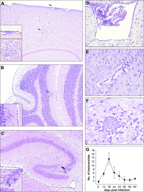
FIG. 4.
Histopathological lesions in brains of MCMV-infected newborn mice. CV staining of different brain regions show acute meningitis (A) and mononuclear cell infiltration in choroids plexus (D) on day 7 p.i., mononuclear cell infiltration in cerebellar cortex (B), perivascular cuffing in hippocampus (C) on day 11 p.i., and edema (E) and glial node (F) in hindbrain on day 14 p.i. Magnification, ×20 (A to E) and ×40 (F and insets). Histopathological lesions were observed in brain parenchyma even at day 80 p.i. (G).

