FIG. 6.
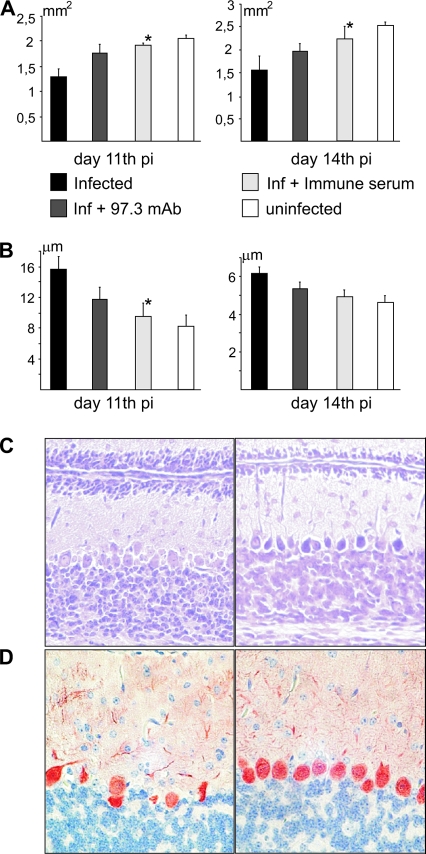
Antiviral antibodies prevent MCMV-induced developmental abnormalities in the cerebella of newborn mice. (A) Cerebellar area in infected and uninfected newborn mice. Mice treated with immune serum on day 5 p.i. show significant increase (*, P < 0.05) in cerebellar area compared to untreated infected mice (days 11 and 14 p.i.). Mice treated with MAb 97.3 on day 5 p.i. show improvement in cerebellar area but without statistically significant difference to control infected mice. (B) EGL thickness in MCMV-infected and uninfected cerebellum. Mice treated with immune serum on day 5 p.i. show significantly decreased (*, P < 0.05) EGL thickness on day 11 p.i. compared to untreated infected mice. Mice treated with MAb 97.3 also have decreased EGL thickness but with no statistical difference relative to control infected animals. (C) EGL thickness in MCMV-infected cerebellum treated with control serum (left) or immune serum (right) on day 12 p.i. is shown in CV-stained sections. Magnification, ×20. (D) Purkinje cell alignment in MCMV-infected cerebellum is modified by treatment with antiviral antibodies. MCMV-infected cerebellum (left panel) and cerebellum of infected, immune serum-treated mice (right panel) are shown. Anti-calbindin MAb was used for immunohistochemical staining. Magnification, ×40.

