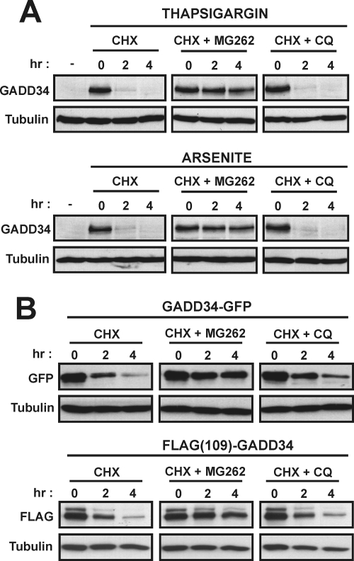
FIG. 4.
Proteasomal degradation of GADD34. (A) SW480 cells exposed to 1 μM thapsigargin or 25 μM arsenite for 5 h to induce GADD34 were treated with CHX (30 μg/ml) either alone or in combination with MG262 (5 μM) or CQ (200 μM), and GADD34 levels were analyzed at selected times by immunoblotting. (B) GADD34 fused at its C terminus to GFP (GADD34-GFP) or containing an internal FLAG epitope, FLAG(109)GADD34, were expressed in HEK293T cells. After CHX treatment to inhibit protein synthesis, degradation of the expressed GADD34 proteins was analyzed by immunoblotting with anti-GFP or anti-FLAG antibodies. Cells were treated with either MG132 or CQ to investigate the role of proteasome or lysosome in GADD34 degradation. Tubulin levels monitored in all immunoblots ensured equal protein loading.

