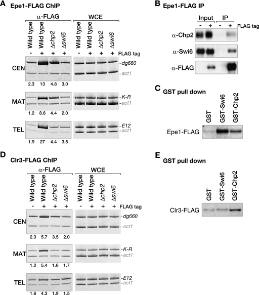
FIG. 4.
Distinct contributions of Swi6 and Chp2 to the heterochromatin localization of Epe1 or Clr3. (A) Epe1 localization to heterochromatic loci is differentially regulated by Swi6 or Chp2. DNA isolated from an anti-FLAG-immunoprecipitated chromatin fraction (α-FLAG) or whole-cell extract (WCE) was used as a template for PCR amplifying centromeric dg660, mat locus K-R, or telomeric E12. The samples were prepared from control nontagged (−FLAG tag) cells or strains expressing Epe1-FLAG (+FLAG tag) from its native promoter. The enrichment ratios of the dg660, K-R, or E12 signals to the act1 signals in the ChIP results are shown beneath each lane. (B) Epe1 efficiently interacts with both Swi6 and Chp2. Anti-FLAG antibodies were used to immunoprecipitate Epe1-FLAG from a strain expressing it (IP). Chp2, Swi6, or Epe1-FLAG was detected by Western blotting with anti-Chp2, anti-Swi6, or anti-FLAG antibodies, respectively. (C) Both Swi6 and Chp2 physically interact with Epe1. Control GST, GST-Swi6, or GST-Chp2 was incubated with whole-cell lysate prepared from cells expressing Epe1-FLAG. Pulled-down Epe1-FLAG was detected by Western blotting using the anti-FLAG antibody. (D) Clr3 localization at heterochromatic loci depends on both Swi6 and Chp2. Association of Clr3-FLAG with the indicated heterochromatic regions was assayed by ChIP as in panel A, using the anti-FLAG antibody. (E) Chp2 has a higher affinity than Swi6 for Clr3. The association of Clr3-FLAG with either Swi6 or Chp2 was assayed as in panel C. α, anti.

