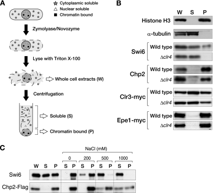
FIG. 6.
Chp2 and Swi6 bind the chromatin-enriched nuclear subfraction in distinct manners. (A) Schematic representation of the chromatin fractionation assay used in the present study. Whole-cell extracts (W) were spun and separated into soluble supernatant (S) and chromatin-enriched pellet (P) fractions. (B) Chromatin fractionation assay in the presence or absence of the histone H3K9 methyltransferase gene, clr4+. Proteins from each fraction were separated by SDS-PAGE, and Swi6, Chp2, Clr3-myc, or Epe1-myc was detected by Western blot analysis. Histone H3 and α-tubulin were examined as controls. (C) Chromatin fractionation assay with various concentrations of salt. The pellet fraction was treated with 0 to 1,000 mM NaCl followed by centrifugation to divide the extract into supernatant and pellet fractions. Swi6 and Chp2-FLAG were detected by Western blotting.

