
Fig. 3.
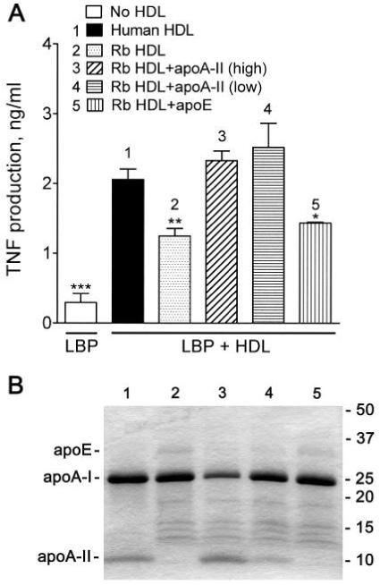
Incorporation of apoA-II restores normal activity to apoA-II-poor nHDL. (A) Rabbit plasma, which contains very low concentrations of apoA-II, was incubated at 37°C for 30 min with 400 μg/ml (3) of purified human apoA-II, the normal apoA-II concentration of human plasma, or with 133 μg apoA-II/ml (4) to allow apoA-II incorporation into HDL; human apoE (5) was used as a control at 50 μg/ml, the normal concentration of apoE in human plasma. The HDL was then isolated by ultracentrifugal flotation. THP-1 cells were stimulated with LPS as described in the caption to Figure 1 in the presence of an inhibitory concentration of LBP (1 μg/ml) and HDL (200 μg protein/ml) isolated from control human (1) or rabbit (2) plasma or HDL from rabbit plasma (3-5) containing the indicated apolipoproteins. Error bars are mean ± SEM of four determinations in two incorporation experiments. Significant differences from the activity of human HDL are denoted * P = 0.0163, ** P = 0.0043, *** P = 0.0001. (B) The above HDL preparations (5 μg protein) were separated on 4-20% SDS-PAGE gradient gels (reducing conditions), and the proteins were stained with Coomassie Blue. The lane numbers refer to the samples described in (A).