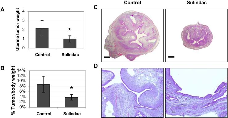
Figure 1. Sulindac inhibits uterine tumorigenesis in the HMGA1a mice.
A, At 29 weeks the uterine tumors from the transgenic mice treated with sulindac were significantly smaller that those of control mice. The bars show the mean uterine weight (grams) ± standard deviation (SD) from 5 mice in each group (*, p = 0.024).
B, In this panel, the uterine tumor weight was divided by the total body weight. Again, tumors in mice treated with sulindac were significantly smaller (*, p = 0.014).
C, Transverse section through the uterine horn from a representative HMGA1a transgenic mouse treated with sulindac (left panels) compared to an untreated mouse (right panels; bars, 500 μm). The uterine horn of the untreated mouse is expanded and filled by an intracavitary stromal proliferation. In contrast, the horn and intracavitary lesion are markedly reduced in size.
D, Higher magnification of the uterine tumors from an HMGA1a mouse treated with sulindac compared to control (bars, 100 μm) demonstrates a polypoid herpercellular and edematous stromal proliferation with intimately associated glandular epithelium in the untreated mouse. In the treated mouse, the lesion is reduced to an attenuated endometrium lacking the polypoid proliferation.

