FIG. 3.
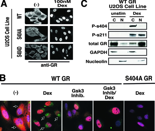
Effects of Ser404 phosphorylation on GR cellular localization. (A) WT-, S404A-, or S404D-GR-expressing U-2 OS cells were treated with 100 nM Dex for 1 h. Cells were then fixed and stained with antibodies directed against human GR. (B) WT- and S404A-GR-expressing U-2 OS cells were treated with Dex (100 nM) and/or the GSK-3α/β inhibitor BIO (5 μM) for 1 h. Cells were then fixed and stained with the nuclear dye DAPI (blue), antibodies directed against human GR (red), and antibodies directed against phospho-S404-GR (green). Arrows indicate phosphorylated GR. All images were captured on a Zeiss LSM 410 laser-scanning microscope. (C) WT-GR-expressing U-2 OS cells were treated with Dex (100 nM) for 1 h before whole-cell lysates were subjected to nuclear and/or cytoplasmic fractionation.

