Fig. 1.
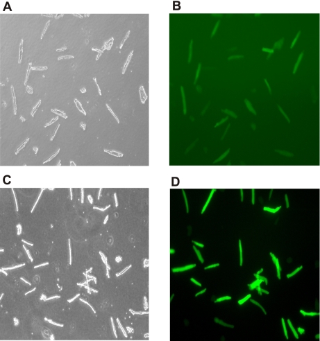
Culture and adenoviral infection of adult mouse myocytes. Cardiac myocytes were isolated from adult congenic phospholemman (PLM) knockout (KO) mice, infected with adenovirus expressing both green fluorescent protein (GFP) and the PLMS68A mutant, and placed in culture as described in methods. A: transmitted light image of adult mouse myocytes after 24 h of culture. B: fluorescent image of A demonstrating the expression of GFP. C: transmitted light image of another adult mouse myocyte culture at 48 h. D: fluorescent image of C. Both B and D are raw images and were taken at the same time using identical excitation light intensities, with the camera set at the same gain but with autoexposure mode. The higher background in B compared with D is due to the longer exposure time because of the much lower GFP fluorescence emission from adenovirus-infected myocytes cultured for 24 h. In contrast, GFP fluorescence emission from infected myocytes cultured for 48 h was much stronger and required a shorter exposure time, as indicated by the lower background.

