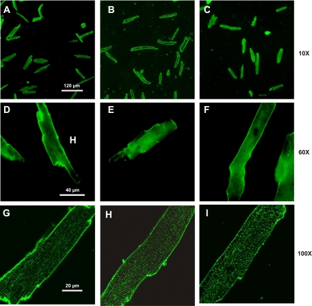
Fig. 2.
Effects of culture on t-tubules in mouse cardiac myocytes. Cardiac myocytes were isolated from wild-type (WT) C57BL/6 mice, plated on laminin-coated coverslips, and cultured for 24–48 h. Myocytes were stained with di-8-ANEPPS and imaged with both conventional wide-field fluorescence (A–F) and confocal (G–I) microscopes. Images acquired with ×10 (A–C), ×60 (D–F), and ×100 (G–I) objectives of myocytes at day 0 (A, D, and G), day 1 (B, E, and H), and day 2 (C, F, and I) of culture are shown.

