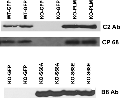
Fig. 4.
Expression of WT PLM and its Ser68 mutants in cultured PLM KO myocytes. Adult cardiac myocytes isolated from congenic PLM KO mice were infected with adenovirus expressing either GFP alone (KO-GFP), GFP + WT dog PLM (KO-PLM), GFP + PLMS68A (KO-S68A), or GFP + PLMS68E (KO-S68E) and cultured for 48 h. Myocytes from WT littermates infected with adenovirus expressing GFP (WT-GFP) and cultured for 48 h were used as controls. Proteins in myocyte lysates (50 μg/lane) were separated by gel electrophoresis and transferred to ImmunBlot polyvinylidene difluoride membranes. Endogenous mouse and WT dog PLM, unphosphorylated and phosphorylated at Ser68, was probed with C2 and CP68 anti-PLM antibodies, respectively, whereas dog PLM Ser68 mutants were detected with B8 anti-PLM antibody. C2 antibody recognizes predominantly the unphosphorylated COOH-terminus of both dog and rodent PLM, and CP68 antibody is specific for PLM phosphorylated at Ser68 (16, 26), whereas B8 antibody recognizes the NH2-terminus of dog but not rodent PLM (18).

