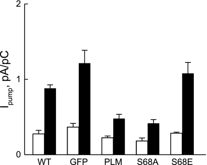
Fig. 7.
Effects of WT PLM and its Ser68 mutants on Na+-K+-ATPase current (Ipump) in cultured PLM KO myocytes. Adult myocytes from congenic PLM KO hearts were infected with adenovirus expressing either GFP alone, GFP + WT PLM (PLM), GFP + PLMS68A (S68A), or GFP + PLMS68E (S68E). After 48 h of culture, Ipump was measured at 18 mM extracellular K+ concentration at 0 mV at 30°C with pipette Na+ concentrations ([Na+]pip) of either 10 mM (open bars) or 80 mM (solid bars). For comparison, Ipump of WT myocytes is shown. At 10 mM [Na+]pip, there were 8 WT, 9 KO-GFP, 10 KO-PLM, 6 KO-S68A, and 7 KO-S68E myocytes. At 80 mM [Na+]pip, there were 5 WT, 7 KO-GFP, 6 KO-PLM, 8 KO-S68A, and 8 KO-S68E myocytes. Two-way ANOVA indicated significant differences between WT and KO-GFP (P ≤ 0.05), WT and KO-PLM (P < 0.0004), WT and KO-S68A (P < 0.001), KO-GFP and KO-PLM (P < 0.002), KO-GFP and KO-S68A (P < 0.003), KO-PLM and KO-S68E (P < 0.003), and KO-S68A and KO-S68E (P < 0.004) myocytes, but not between KO-GFP and KO-S68E (P < 0.8), KO-PLM and KO-S68A (P < 0.8), and WT and KO-S68E (P < 0.3) myocytes.

