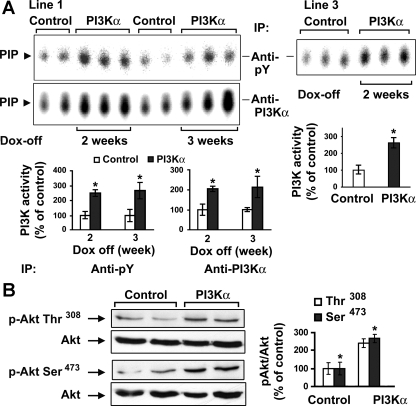
Fig. 2.
Activation of cardiac phosphatidylinositol 3-kinase (PI3K) and Akt in the conditional transgenic mice. A: PI3K activity in 2 double transgenic mouse lines. Three-month-old male line 1 double transgenic littermates were maintained with doxycycline (Dox) water (control; n = 4) or regular drinking water (PI3Kα; n = 6) for 2 or 3 wk (left). Similarly, line 3 mice were maintained with Dox water (control; n = 3) or regular drinking water (PI3Kα; n = 3) for 2 wk (right). Cardiac PI3K activity was assessed with in vitro lipid kinase assay. Phosphoinositide 3-phosphate (PIP) is the phosphorylated end-product. Each bar graph shows the densitometric scanning results from 2 individual experiments. Data are expressed as means ± SE of percent change in PI3K activity relative to that of control. *P < 0.05 vs. control. B: Western blotting was performed on cardiac tissue lysates from male control (n = 2) and PI3Kα (2 wk off Dox; n = 2) line 1 mice using phospho (p)-specific antibodies against Akt (Ser-473 and Thr-308) and an antibody against total Akt. The bar graphs show results of densitometric measurement of each phospho-specific antibody normalized with total antibody from 3 separate experiments. *P < 0.05 vs. control. pY, phospho-tyrosine; IP, immunoprecipitation.

