Fig. 3.
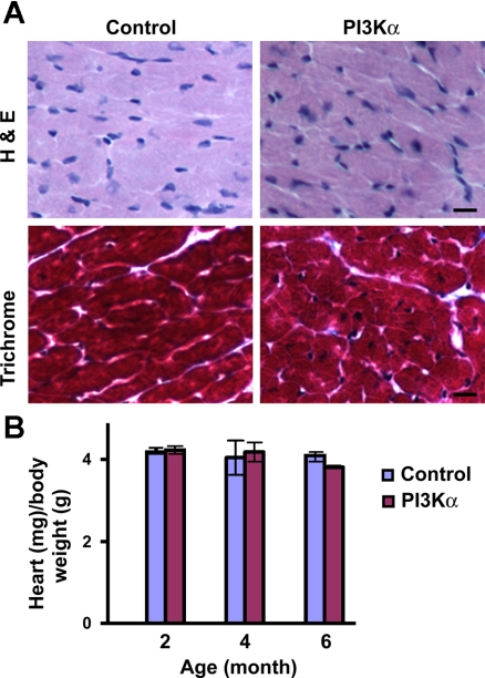
Absence of cardiomyopathy and hypertrophy following temporal (2 wk) overexpression of cardiac-specific PI3Kα. A: histological analysis of heart sections from 3-mo-old male PI3Kα and control littermate. A, top: hematoxylin and eosin (H & E) staining. A, bottom: Masson trichrome staining. Bars represent 10 μm. B: heart weight-to-body weight ratio in PI3Kα and control littermates. Mice were 2 (n = 8), 4 (n = 10), or 6 (n = 8) mo old before initiation of cardiac PI3Kα overexpression.

