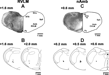
Fig. 7.
Histological identification of microinjection sites. A: coronal section of the medulla at a level 1.6 mm rostral to the calamus scriptorius (CS) showing a typical RVLM site marked with India ink (100 nl) where NBQX and d-AP7 microinjections were made. The center of the spot was 1.8 mm lateral to the midline and 2.7 mm deep from the dorsal medullary surface. B: drawings of coronal sections at a level 1.6 and 2.0 mm rostral to the CS. In this and other panels, microinjection sites are shown as dark spots; each spot represents a site in one animal. C: coronal section of the medulla at a level 0.6 mm rostral to the CS showing a typical nAmb site marked with India ink where NBQX and d-AP7 or hydroxysaclofen and gabazine microinjections were made. The center of the spot was 1.9 mm lateral to the midline and 2.3 mm deep from the dorsal medullary surface. D: drawings of coronal sections at a level 0.2, 0.5, and 0.6 mm rostral to the CS. Amb: nucleus ambiguus; AP: area postrema; CVLM: caudal ventrolateral medullary depressor area; ECu: external cuneate nucleus; NTS: nucleus tractus solitarius; Py: pyramids; Sp5: spinal trigeminal tract; 10: dorsal motor nucleus of vagus.

