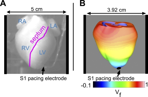
Fig. 1.
A: view of the anterior rabbit heart in the experimental setup. The locations of the right atrium (RA), left atrium (LA), right ventricle (RV), left ventricle (LV), and septum are marked. The S1 pacing bipolar electrode is located near the apex, and the shock electrodes are positioned at the right and left sides of the perfusing chamber. B: schematic of the three-dimensional model of the rabbit ventricles with computed optical fluorescence signal (Vf) shown during the repolarization of an S1-paced beat. S1 stimuli are delivered via the apical pacing electrode, as indicated by the black arrow; field shocks are administered via plate electrodes located on either side of the ventricles in the chamber (as represented by the vertical black bars).

