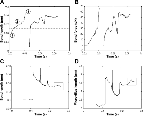
Fig. 6.
Sample length and force traces for individual bonds and microvilli of modeled PMNs rolling over P-selectin substrates. Bond lengths (A) and bond forces (B) were tracked for an individual bond attached to a rigid microvillus as a function of time. Bond lengths (C) and microvillus lengths (D) were also recorded for the case of a deformable microvillus. Whereas the microvillus length followed the same pattern as the bond initially, it is noteworthy that microvillus length increased, whereas the bond length remained nearly constant, later in the simulation (indicated by boxes in C and D). The P-selectin site density was 15 molecules/μm2, and the cell was subjected to a uniform shear rate of 10 s−1. The membrane stiffness was 3 dyn/cm.

