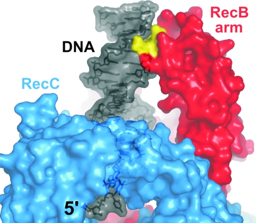
FIG. 10.
The RecB arm. Shown is a close-up view of the “arm” structure (red) formed by an auxiliary subdomain (subdomain 1B) of RecB. The arm contacts duplex DNA ahead of the translocating enzyme. A conserved patch of residues in close proximity to the minor groove of the DNA substrate (black semitransparent model) is shown in yellow. The RecC protein is in the foreground (blue). The 5′ ssDNA tail is pointing toward the viewer through a hole in the RecC protein.

