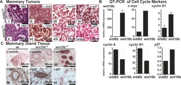
Figure 1.
p27KIP1 protein, but not mRNA, is reduced in Wnt10b transgenic mammary tissue and elevated in Wnt10b-null mammary tissue. (A) Five-micrometer paraffin sections from Wnt10bTG (panels i–iv) and MMTV-ErbB2 transgenic (ErbB2TG) (panels v–viii) mammary tumor tissue were stained with H&E (panels i,v) to show morphological features. In parallel sections, protein expression was characterized by IHC (brown): β-catenin (panels ii,vi), cyclin D1 (panels iii,vii), and p27KIP1 (panels iv,viii). Sections were counterstained with nuclear fast red (red) to reveal nuclei. (B) RNA expression profiling (see Supplemental Fig. S2) and QPCR expression analysis was carried out on cDNA from similar tumors. Transcripts were quantified, and values were normalized to 18S rRNA. (C) p27KIP1 (panels i–iii) and WNT10b (panels iv–vi) protein expression was examined in 7-wk-old mammary tissue from wild-type (wt), Wnt10b-null (Wnt10b−/−), and Wnt10bTG mice. Five-micrometer paraffin sections were stained as described above. Bars indicate scale in micrometers.

