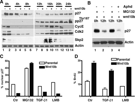
Figure 2.
Mammary cell lines expressing Wnt10b show accelerated turnover of p27KIP1 protein in late G1/early S phase, prior to the induction of SKP2. Turnover is blocked by inhibitors of proteasome function but not by inhibitors of CRM-1 nuclear export. NMG and NMG stably expressing Wnt10b (NMG-Wnt10b) cells were synchronized in G1 phase and released by replating. Cell extracts were prepared at the indicated times. (A) Lysates were analyzed by immunoblotting and probed with the indicated antisera. (B) NMG-Wnt10b cells were synchronized at G1 phase. Following release, cells were cultured in the presence or absence of the proteasome inhibitor MG132 (10 μM) or the S-phase blocker aphidicolin (1 μg/mL). Twelve-hour samples were immunoblotted as described above. (C) Nuclear p27KIP1 levels were measured in NMG vector (parental) and NMG-Wnt10b cells following exposure to proteasome blocker MG132 (10 μM), TGFβ (2.5 ng/mL), and LMB (1 μg/mL). Cells were synchronized at G1 phase and released for 12 h, and p27 immunofluorescence was measured quantitatively using an LSC laser cytometer. (D) BrdU incorporation into cells from C was detected by immunofluorescence, and signal was measured quantitatively using an LSC laser cytometer. Experiments were conducted as triplicates. Error bars represent SDM.

