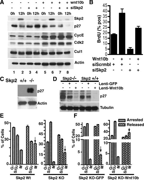
Figure 4.
Wnt10b stimulates turnover of p27KIP1 protein in the absence of SKP2 and promotes M-phase entry in Skp2−/− embryonic fibroblasts following synchronization. NMG-control and NMG-Wnt10b cell lines were transfected with pSuppressorNeo vector generating synthetic siRNA for Skp2. (A) Cells were harvested at 0 h and 12 h post-release. Whole-cell lysates were analyzed for expression of SKP2, p27, cyclin E, CDK2, Cullin 1, and β-actin. (B) Cells from A were subsequently pulsed for 15 min with BrdU to label S-phase entry and then fixed at 12 h post-release. BrdU incorporation was assessed by immunofluorescence and quantified by LSC. (C) MEFs from Skp2+/+ and Skp2−/− mice were synchronized in early S phase in the presence of aphidicolin (1 μg/mL). Whole-cell lysates were analyzed for p27KIP1 by immunoblotting. (D) Skp2−/− and Skp2+/+ MEFs were stably transduced with lentivirus expressing GFP or Wnt10b and synchronized with aphidicolin. Whole-cell lysates were immunoblotted to identify p27KIP1 and tubulin. (E) Wild-type and Skp2−/− MEFs from C were arrested in S phase by aphidicolin treatment, released for 24 h and then analyzed for M-phase entry using a nuclear morphology protocol established for the LSC (Luther and Kamentsky 1996). (F) Skp2−/− MEFs cells (±Wnt10b) from D were analyzed for M-phase entry after release. Arrows identify the fraction of M cells 8 h after release into S phase. Experiments were conducted in triplicate, and error bars represent SDM.

