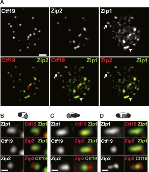
Figure 3.
Zip2 and centromere configurations in short, linear Zip1 stretches. (A) Spread nucleus from wild type at early zygotene stained with antibodies to Ctf19, Zip2, and Zip1. The arrows and arrowheads indicate Zip1 stretches containing one or two Zip2 foci, respectively. Bar, 2 μm. (B–D) Examples of the most common configurations of Zip2 and Ctf19. In the diagram at the top of each panel, unfilled ovals indicate Zip1; gray circles indicate centromeres, and black regions indicate Zip2. Bar, 0.125 μm.

