


Fig. 2.
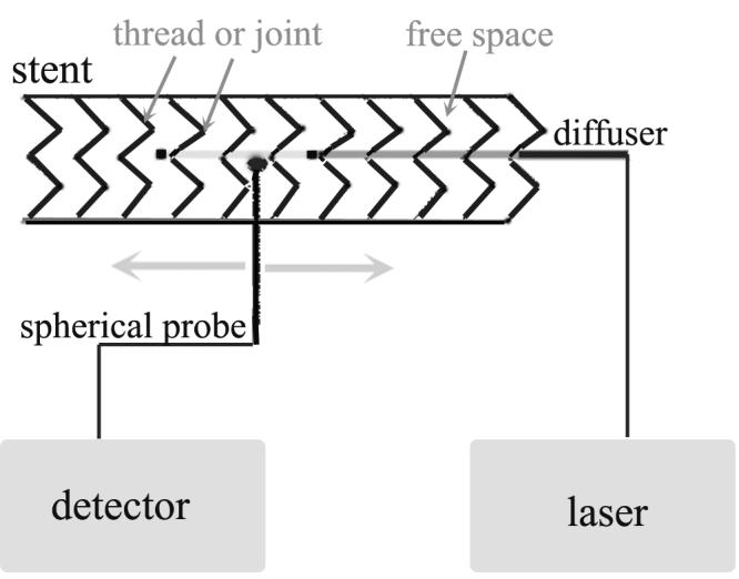
Experimental setup of point measurement. The light source (spherical probe or diffuser – showing in the diagram) was placed inside the stent. The measurement probe (spherical probe) was pointed to the metal thread, joint or free space in a touch mode (0 mm) or 1 - 2 mm away from the outer surface in the presence or absence of the tissue specimen (shown in the diagram).