
Figure 1. Activation of essentially all CD4+Vβ8+ T cells in mouse spleen two hours after injection of SEB.
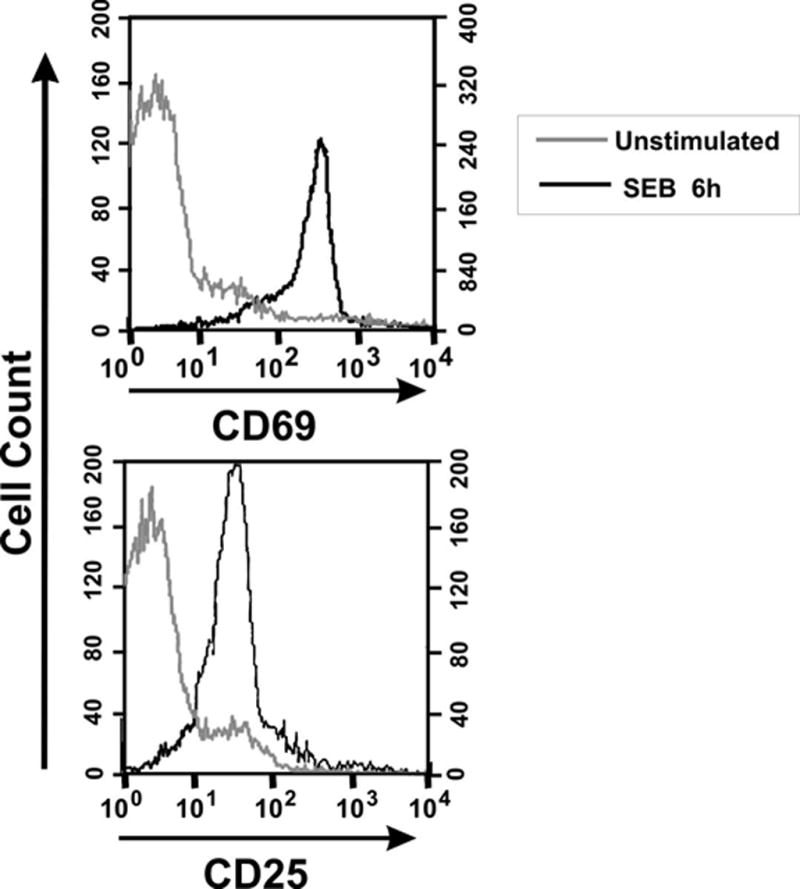
BALB/c mice were injected with 50 μg of SEB in PBS (SEB 6h) or with PBS alone (unstimulated), euthanized 2 h later and their splenocytes evaluated by flow cytometry. Histograms depicting relative expression levels of CD69 and CD25 are gated on CD4+Vβ8+ lymphocytes. Greater than 95% of CD4+Vβ8+ T cells were activated.