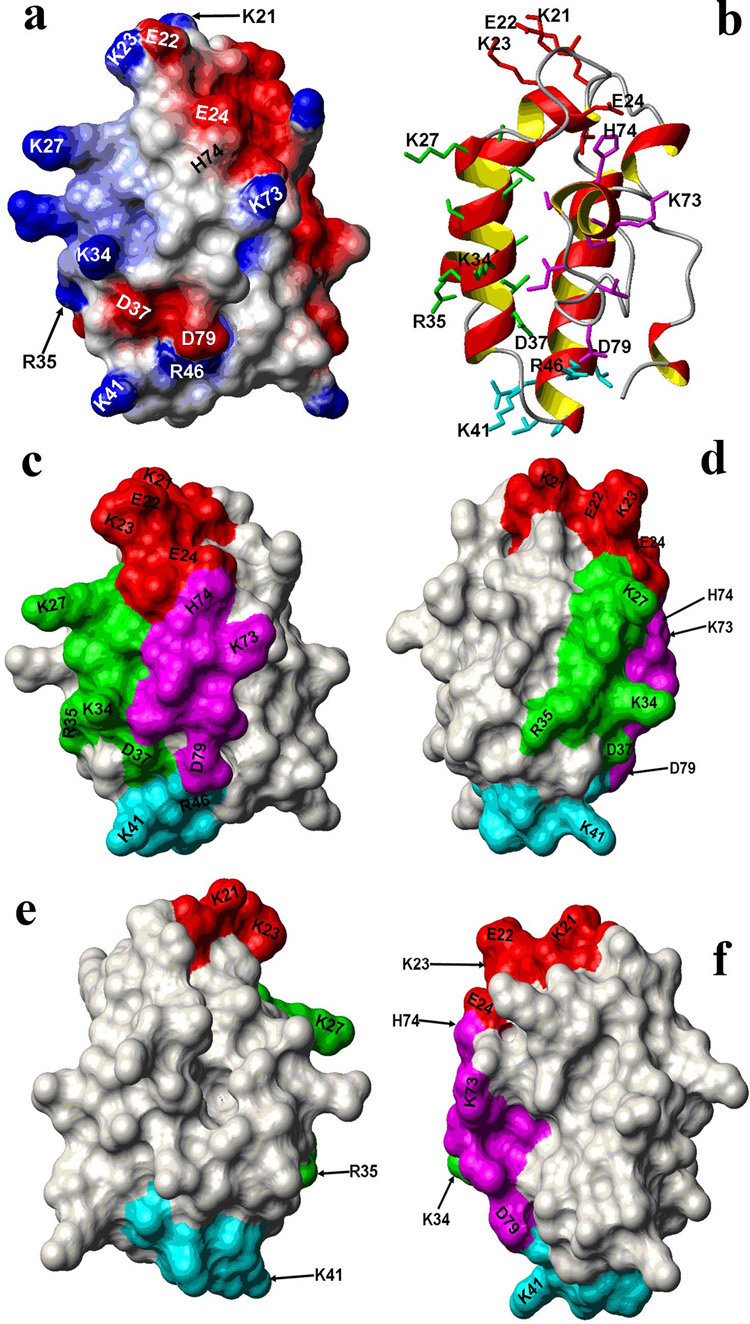
Figure 4.
Linear epitopes map to a hydrophilic, lysine rich face of the 3D-model of the weed pollen allergen Par j 1. Charged residues on the surface of linear epitopes are labeled. a) surface electrostatic potential, b) ribbon plot showing the surface exposed side chains of the linear epitopes, colored to indicate the different epitopes; c–f: rotations around the y-axis, starting from the orientation of panel a, showing the epitopes colored coded as in b). This face is positively charged while the back side is predominantly negative.

