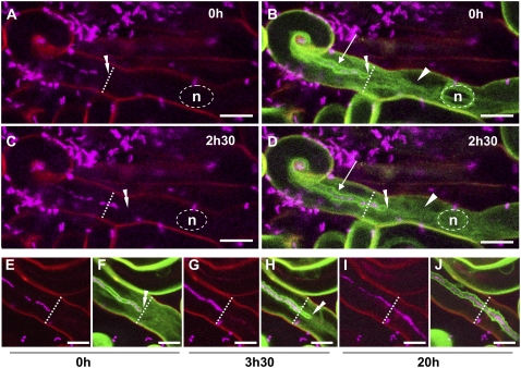
Figure 2.
Labeling of the IT interface with GFP-tagged AtPIP2;1 aquaporin. Growing ITs in two different root hairs of sunn plants expressing the AtPIP2;1-GFP fusion. Confocal images A to J (z axis projections of serial optical sections) show the cCFP fluorescence labeling the rhizobia (magenta) and images B, D, F, H, and J the additional fluorescence of the AtPIP2;1-GFP fusion (green). Cell wall autofluorescence in the confocal images is shown in red. The transverse dashed line indicates the initial position of the IT tip (0 h time point) in each image. A to D, Recently initiated IT (4 d postinoculation) observed at two successive time points (0 h [A and B] and 2.5 h [C and D]). In addition to the plant cell membrane, the entire IT membrane (arrows) is labeled by the aquaporin-GFP fusion (B and D). As in Figure 1, the single file of aligned rhizobia within the IT is interrupted by a number of gaps (A and C). In addition, the aquaporin-GFP label reveals that there is a space between the leading bacteria in the file and the growing tip of the IT (double arrowheads) for both time points (B and D). The weak GFP labeling surrounding the nucleus (n) and within the cytoplasmic bridge (arrowheads) is probably indicative of an intracellular pool of the aquaporin fusion protein. E to J, Progressive growth and rhizobial colonization of an IT (5 d postinoculation) observed at three successive time points in a root hair (0 h [E and F], 3.5 h [G and H], and 20 h [I and J]). Initially, the leading bacterium of the discontinuous file is very close to the tip of the IT (E and F); 3.5 h later the tip has grown several micrometers further down the hair, but the leading bacterium has not moved from its original position (G and H), thus creating a space behind the tip. At the same time, most of the initial gaps within the bacterial file have been filled. Finally, the initial smooth appearance of the IT interface (F) has started to become uneven at the 3.5-h time point (H). Twenty hours later (I and J), the IT has progressed down the shaft of the root hair, and displays the characteristic uneven surface of a mature IT. Note that, as in Figure 1, gaps are still present within the bacterial file of the mature IT, and that certain stretches of the file appear to have doubled. The nucleus of the cell is not visible in any of these images, but in all cases was positioned ahead of the growing thread (data not shown). Bars = 10 μm.

