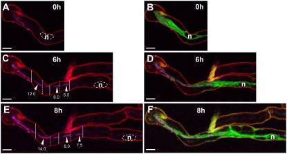
Figure 3.
Bacteria colonize ITs by division and movement. Three successive stages (0, 6, 8 h) of the growth of an IT in a root hair of a wild-type M. truncatula transgenic line expressing the ER-targeted GFP-HDEL (2 d postinoculation). Confocal images A to F (z axis projections of serial optical sections) show the cCFP fluorescence labeling the rhizobia (magenta) and images B, D, and F the additional fluorescence of the GFP-HDEL fusion (green). Cell wall autofluorescence is shown in red. A and B, Early infection stage (0 h). Two short ITs have initiated in one curled root hair. C and D, One of the ITs has elongated and the second has aborted (6 h). Note the bacteria-free spaces separating short files of bacteria within the growing IT. The alternating continuous and dashed vertical lines (C) indicate the positions of the leading bacteria of five contiguous bacterial files separated by spaces. The numbers indicate the length (μm) of three of these bacterial files (arrowheads). E and F, IT has further elongated within the root hair (8 h). The vertical continuous and dashed lines indicate the new positions of the leading bacteria of each of the five bacterial files marked in C. The bacteria have progressed down the thread mainly due to the collective movement of the files. Cell divisions have also taken place in the files, as indicated by their increased length (μm). Bars = 10 μm. n, Nucleus.

