Figure 8.
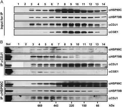
Analysis of complexes formed by HSP90C, HSP70B, CDJ1, and CGE1. A, Size distribution of HSP90C, HSP70B, CDJ1, and CGE1 complexes. Soluble proteins were extracted from Chlamydomonas and protein complexes, after stabilization by crosslinking with 2 mm DSP, were separated by gel filtration. Aliquots of collected fractions were separated on a 7.5% to15% SDS-PAGE and analyzed by immunoblotting using antibodies against HSP90C, HSP70B, CDJ1, and CGE1. B, Analysis of HSP90C- and CDJ1-containing complexes. Gel filtration fractions containing DSP-crosslinked protein complexes were incubated with protein-A sepharose coupled to antibodies against CDJ1 and HSP90C. An aliquot of soluble proteins (representing 3.8% of the input to gel filtration) and precipitated proteins were analyzed by immunoblotting as in A.

