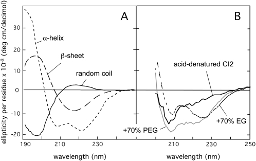
Figure 2.
Reference CD spectra of the common types of secondary structure and random coil conformations. A, Pure α-helical structure (dotted line), pure β-sheet structure (dashed line), and random coil (solid line). B, The acid-denatured state of the globular protein CI2 in pure buffer and in 70% EG and PEG. Notably, the structural induction of CI2 by EG and PEG is not along the normal folding pathway but seems to involve nonnative interactions (Silow and Oliveberg, 2003).

