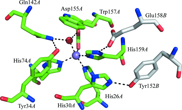
Figure 3.
The structure of the active site of MnSOD-3 showing the hydrogen-bonding network viewed from the approximate direction of substrate access. The manganese and the hydroxyl ions are shown as magenta and red spheres, respectively. Residues from different subunits, which form a dimer, are colored as in Fig. 2 ▶(b). This figure was produced using PyMOL (DeLano, 2008 ▶).

