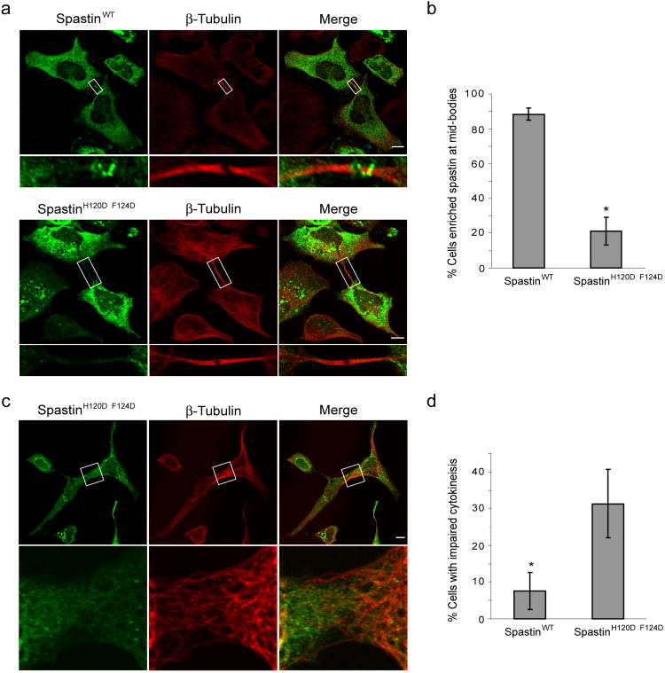
Figure 5. Spastin MIT domain mutant protein shows decreased enrichment at midbodies and alters cytokinesis.
(a) HeLa cells expressing wild-type (WT) Myc-spastin (green; top panels) or else Myc-spastinH120D/F124D (green; bottom panels) were co-immunostained for Myc-epitope and β-tubulin. Myc-spastinH120D/F124D shows decreased enrichment at midbodies, as identified by β-tubulin. Boxed areas are enlarged in the lower panels. Bar, 10 μm. (b) Quantification of enrichment of wild-type versus MIT mutant spastin expressing cells (n=3; 100 cells per experiment; ±SD). *p=0.001. (c) HeLa cells expressing Myc-spastinH120D/F124D (green) exhibited impaired cytokinesis, with microtubules (red) often maintaining a connection between cells. The boxed area is enlarged in the lower panels. Bar, 10 μm. (d) Quantification of cytokinesis impairment, as defined by the persistence of cellular interconnections, in wild-type Myc-spastin versus Myc-spastinH120D/F124D expressing cells (n=3; 100 cells per experiment; ±SD). *p<0.05.

