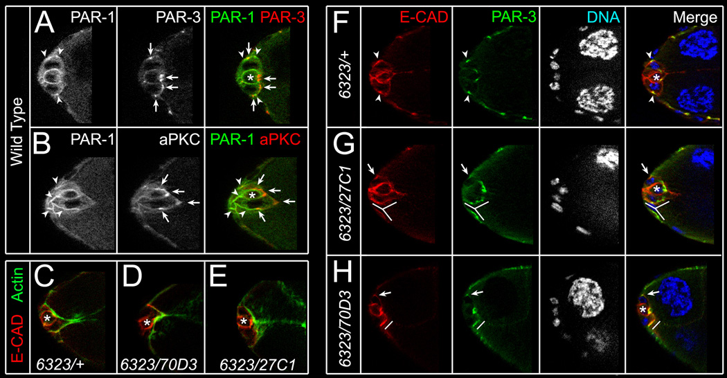
Figure 3. PAR-1 regulates localization of PAR-3 and E-CAD between border and follicle cells.
(A–B) Stage 9 wild-type border cells stained for PAR-1 (green, arrowheads) and PAR-3 (red, arrows; A) or aPKC (red, arrows; B). PAR-1, localized to basolateral cell membranes, is largely non-overlapping with apical PAR-3 and aPKC. (C–E) Stage 9 control (C) and viable par-16323/par-170D3 (D) or par-16323/par-127C1 (E) mutant border cell clusters stained for phalloidin (green) to visualize F-actin and E-cadherin (E-CAD; red) to label cell membranes. F-actin localizes normally. (F–H) Stage 9 control (F) and viable par-16323/par-127C1 (G) or par-16323/par-170D3 (H) mutant border cell clusters stained for E-CAD (red), PAR-3 (green) and DAPI to visualize nuclei (white/blue). (F) E-CAD and PAR-3 co-localize at foci between border cells and follicle cells (arrowheads; F). (G, H) In par-1 mutant egg chambers, E-CAD and PAR-3 are less enriched at foci (arrows; G, H), found in multiple foci (lines; G), or are broadly enriched (lines; H). Single optical sections shown in all panels; polar cells (*).

