
Figure 3.
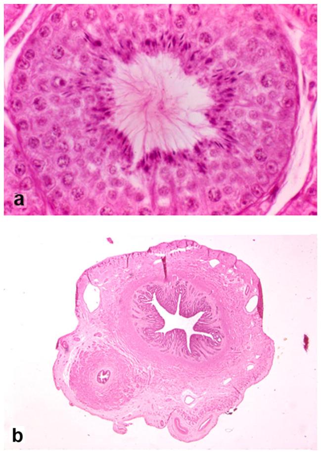
Selected histological features of the reproductive tract in PMDS male A8, age 6 years (hematoxylin and eosin stain): a) seminiferous tubule of left scrotal testis showing normal stages of spermatogenesis (original magnification x400), b) cross section of uterine horn and adjacent vas deferens in parallel which, except for their presence together, have normal characteristics (original magnification x250).