
Figure 5.
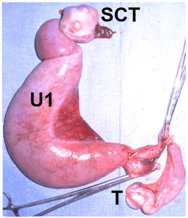
Sequelae to canine PMDS and cryptorchidism. The excised uterine horns, cryptorchid testis and scrotal testis from an aged PMDS male are shown. The uterine horn (U1) attached to the cryptorchid testis is grossly dilated due to pyometra. The remaining uterine horn, attached to the scrotal testis (T), is mildly dilated. Nodules in the markedly enlarged cryptorchid testis were identified by histology as Sertoli cell tumor (SCT).