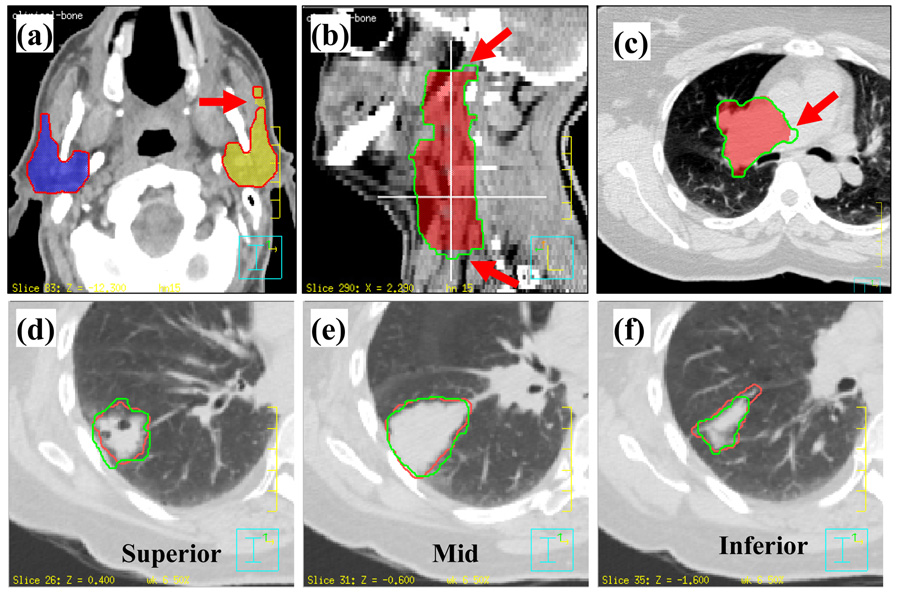
Fig. 4.
Examples of differences between the automatically propagated contours and physician-drawn contours. (a) Deformed contour is separated from the left parotid; (b) the two ends disagree with physician’s contours; (c) contours did not deform in the low contrast region; (d) and (f) manually drawn contours did not agree at the two ends of a structure; (e) manually drawn contours agreed at the middle of a structure. (a),(b),(c) Colorwash: physician’s manually drawn contours; contour: deformed contours. (d),(e),(f) Red: manual contours; green: deformed contours.

