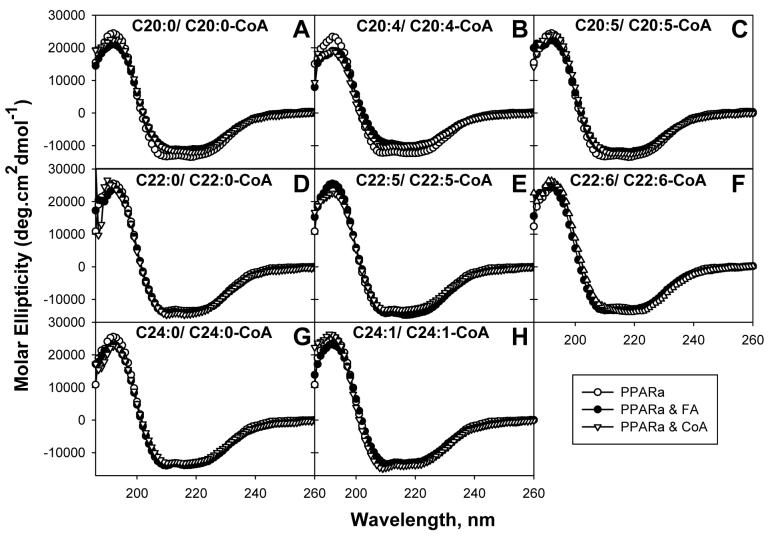
FIG. 6. Effect of VLCFA and VLCFA-CoA binding on PPARαΔAB conformation: Circular dichroic (CD) spectra.
Far ultraviolet (UV) circular dichroic (CD) spectra of PPARαΔAB in the absence or presence of added ligand were obtained as described in the “Experimental Procedures”. Panel A, CD spectrum of PPARαΔAB in the absence (empty circles) and presence of added ligand: arachidic acid (filled circles) or arachidoyl-CoA (empty triangles); panel B, in the presence of arachidonic acid (filled circles) or arachidonoyl-CoA (empty triangles); panel C, in the presence of eicosapentaenoic acid (filled circles) or eicosapentaenoyl-CoA (empty triangles); panel D, in the presence of behenic acid (filled circles) or behenoyl-CoA (empty triangles); panel E, in the presence of docosapentaenoic acid (filled circles) or docosapentaenoyl-CoA (empty triangles); panel F, in the presence of docosahexaenoic acid (filled circles) or docosahexaenoyl-CoA (empty triangles); panel G, in the presence of lignoceric acid (filled circles) or lignoceroyl-CoA (empty triangles); panel H, in the presence of nervonic acid (filled circles) or nervonoyl-CoA (empty triangles). Each spectrum represents an average of 10 scans for a given representative spectrum from three replicates.

