
Figure 17.
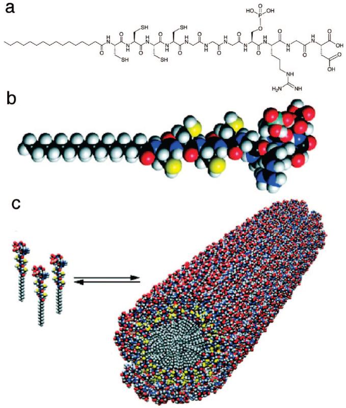
(a) Chemical structure of the PA, consisting of a hydrophobic alkyl tail; four cysteine residues that when oxidized may form disulfide bonds to polymerize the self-assembled structure; a flexible linker region of three glycine residues to provide the hydrophilic head group flexibility from the more rigid cross-linked region; a single phosphorylated serine residue that was designed to interact strongly with calcium ions and help direct mineralization of HA; and the cell adhesion ligand RGD. (b) Molecular model of the PA showing the overall conical shape of the molecule going from the narrow hydrophobic tail to the bulkier peptide region. (c) Schematic showing the self-assembly of PA molecules into a cylindrical micelle. Reprinted with permission from Science (http://www.sciencemag.org), ref 79. Copyright 2001 AAAS.