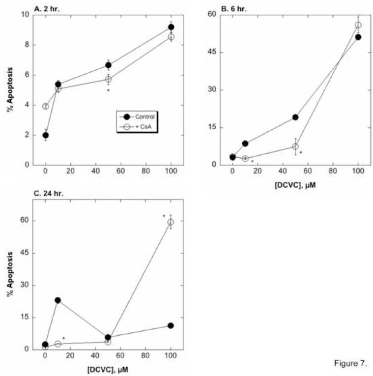
Figure 7. Effect of CsA on DCVC-induced apoptosis.
hPT cells grown in collagen-coated, 6-well plates were incubated for 4 or 24 hr with the indicated concentrations of DCVC in the absence or presence of 0.5 nmol CsA/mg protein. Cells were stained with propidium iodide and cell cycle status was analyzed by flow cytometry and FACS analysis. Results are the percentage of cells undergoing apoptosis (sub-diploid) and are means ± SEM of measurements from 4 separate cell cultures, each deriving from a different kidney. Where not visible, error bars were within the bounds of the data point. *Significantly different (P < 0.05) from the corresponding control cells.

