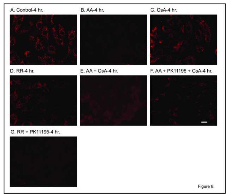
Figure 8. Effect of mitochondrial inhibitors on mitochondrial membrane potential.
Confluent, primary cultures of hPT cells were grown on collagen-coated, glass cover slips in 35-mm tissue culture dishes and were incubated for 4 hr with media (= Control) or mitochondrial inhibitors, added singly or together, as indicated, at the following concentrations: 1 μM AA, 0.5 nmol CsA/mg protein, 30 μM RR, and 120 μM PK11195. JC-1 fluorescence was measured by confocal microscopy assessing the emission of punctate red (∼590 nm) in polarized mitochondria using 488-nm excitation. Red fluorescence is shown. Polarized mitochondria are indicated by yellow-red punctate staining whereas depolarized mitochondria exhibit diffuse or no staining. White bars = 5 μm.

