
Figure 4.
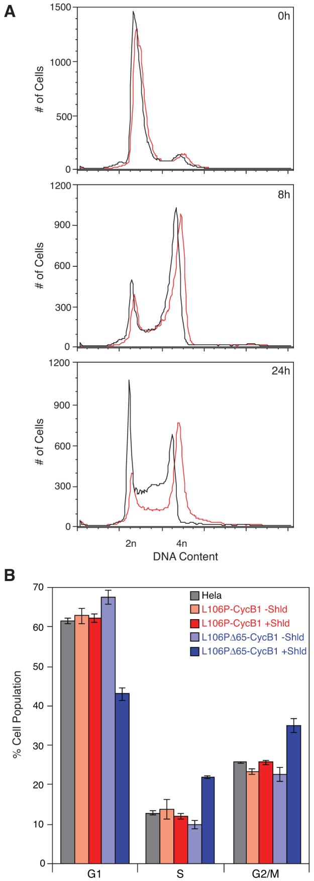
HeLa cells expressing Cyclin B1 constructs were treated with Shield-1 or mock-treated following release from cell cycle arrest and incubated for the indicated periods of time. Cells were then analyzed by flow cytometry for DNA content. (A) Analysis of Shield-1-treated (red) and mock-treated (black) cells expressing L106P-Δ65Cyclin B1 at 0h, 8h, and 24h post-release from arrest. (B) Analysis of Shield-1-treated (faded colors) and mock-treated (solid colors) cells expressing L106P-Cyclin B1 (red) or L106P-Δ65Cyclin B1 (blue) 12 hours after release from arrest. The experiment was performed in triplicate (± s.d).