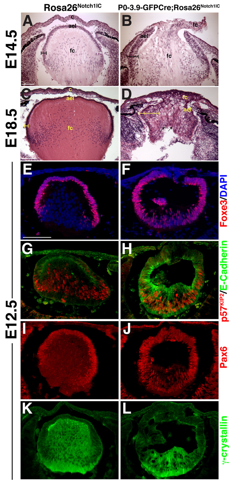
Figure 3. Constitutive expression of Notch signaling alters lens growth and differentiation.
(A–D) Histological sections from the P0-3.9-GFPCre;Rosa26Notch1IC eyes show a thickening and multilayering of the AEL (indicated by brackets), as well as loss of a definitive cornea. At E18.5 (C,D), radially-aligned nuclei are observed in the P0-3.9-GFPCre;Rosa26Notch1IC lens (arrows in D). ael–anterior epithelial layer, c–cornea, fc–presumptive fiber cells. (E,F) Foxe3, (G,H) E-Cadherin, and (I,J) Pax6 expression inappropriately persist in E12.5 posterior P0-3.9-GFPCre;Rosa26Notch1IC lenses, indicating delayed primary fiber cell differentiation, accompanied by a (K,L) highly reduced γ-crystallin domain. (G,H) p57Kip2 initiates normally in the P0-3.9-GFPCre;Rosa26Notch1IC posterior lens in E-Cadherin-positive cells. Bar in A,C,E = 100 microns, anterior is up in A–L.

