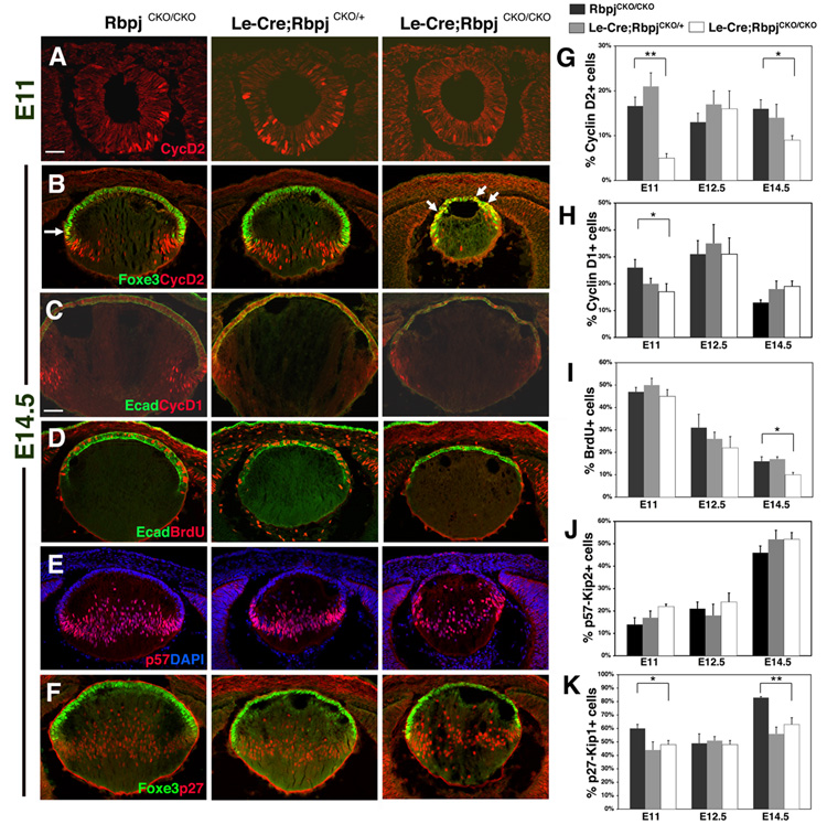
Figure 6. Lens progenitors are reduced in conditional Rbpj mutant lenses.
(A,B) Cyclin D2+ cells are decreased in E11 and E14.5 Le-Cre;RbpjCKO/CKO mutant lenses. At E14.5, Cyclin D2 is improperly expressed throughout the AEL, determined by Foxe3 co-expression (arrows). (C) Cyclin D1+ cells are decreased in E14.5 Le-Cre;RbpjCKO/CKO lenses, especially around the exit point of the transition zone. (D) BrdU+ S-phase cells are decreased in E14.5 Le-Cre;RbpjCKO/CKO eyes. (E) p57Kip2 expression is unaltered in Le-Cre;RbpjCKO/CKO lenses at E11, E12.5 and E14.5. (F) p27Kip1 cells are decreased in Le-Cre;RbpjCKO/CKO lenses, especially at the transition zone in Foxe3+ cells (not shown). (G–K) Quantification of proliferation markers as indicated. Bar in A = 40 microns, in C = 20 microns; anterior is up in A–F. Bar graphs show mean + s.e.m. * = P<0.05. ** = P<0.01.

