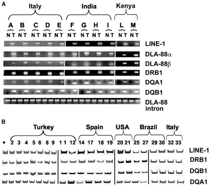
Figure 1. Specific LINE-1/c-myc and DLA Haplotype Genetic Markers for CTVT Detected by Specific PCR Amplification.
(A) For each of 11 dogs (A–M), fresh normal and tumor samples are indicated as N and T, respectively. The panel is assembled from three separate gels visualized by ethidium bromide. The invariant DLA-88 intron sequence serves as a positive control for each of the 22 specimens.
(B) PCR amplification of DNA using Cy5-labeled forward primers from 21 microdissected tumor cells from paraffin-embedded specimens. The panel is assembled from four separate gels.

