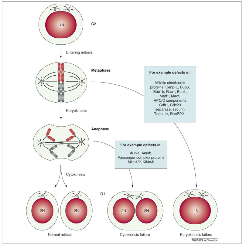
Figure 4.
Tetraploidization by mitotic failure. Normally a 4N cell in G2 enters mitosis, aligns its chromosomes in the metaphase plate and equally distributes the DNA over two nuclei (karyokinesis) and subsequently two daughter cells (cytokinesis). When karyokinesis fails, cells with 4N content are observed. When cytokinesis fails, the DNA is divided into two nuclei that remain within one cell. Mitotic genes that could have a role in failure of karyokinesis and cytokinesis are indicated (blue boxes). Note that cells undergoing karyokinesis and cytokinesis both inherit two centrosomes that could lead to abnormal spindles and chromosome mis-segregation during the next round of division (Box 1).

