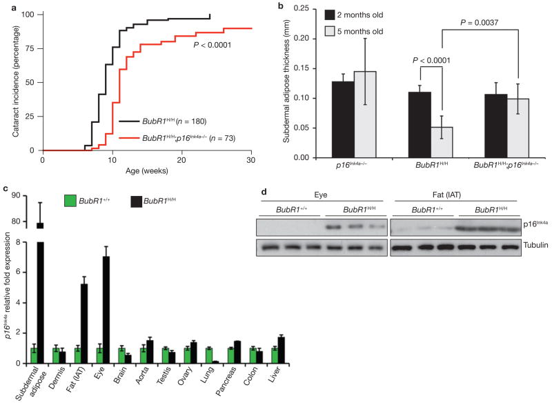
Figure 3. p16Ink4a disruption attenuates selective progeroid features of BubR1 hypomorphic mice.
(a) Incidence and latency of cataract formation in BubR1H/H and BubR1H/H;p16Ink4a−/− mice as detected by the use of slit light after dilation of eyes. The curves are significantly different (P < 0.0001, log-rank test). We note that no wild-type or p16Ink4a−/− mice developed cataracts during this observation period. (b) Subcutaneous adipose layer thickness of p16Ink4a−/−, BubR1H/H and BubR1H/H;p16Ink4a−/− mice at 2 and 5 months of age. Data are mean ± s.d. (n = 4 male mice for each age per genotype). A two-tailed Mann-Whitney test was used for statistical analysis. (c) qRT–PCR analysis for relative expression of p16Ink4a in a variety of 2-month-old tissues from BubR1H/H and wild-type mice. Values were normalized to GAPDH, and relative fold is to 2-month-old wild-type samples. Data are mean ± s.d. (n = 3 male mice for each tissue, with triplicate measurements taken). (d) Western blots of eye and fat extracts from 2-month-old BubR1+/+ and BubR1H/H mice probed with anti-p16Ink4a antibody. Anti-tubulin antibody served as loading control. Uncropped images of the scans are shown in Supplementary Information, Fig. S6b, c.

