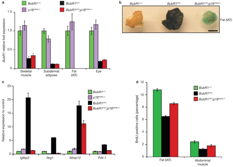
Figure 4. p16Ink4a induction in BubR1H/H mice promotes cellular senescence.
(a) Relative expression of BubR1 in gastrocnemius, subdermal adipose, fat deposits and eyes from 2-month-old wild-type, p16Ink4a−/−, BubR1H/H and BubR1H/H;p16Ink4a−/− mice as determined by qRT–PCR. Values were normalized to GAPDH. Relative fold is to 2-month-old wild-type samples. Data are mean ± s.d. (n = 3 male mice per genotype, with triplicate measurements taken per sample). We note that ablation of p16Ink4a was unable to increase the amount of BubR1 present in either wild-type or BubR1 hypomorphic mice. (b) IAT of 5-month-old wild-type, BubR1H/H and BubR1H/H;p16Ink4a−/− mice stained for SA-β-galactosidase activity. Scale bar is 2 mm. (c) Relative expression of senescence markers in gastrocnemius muscles of 2-month-old wild-type, p16Ink4a−/−, BubR1H/H and BubR1H/H;p16Ink4a−/− mice analysed by qRT–PCR. Data are mean ± s.d. (n = 3 male mice per genotype). Values were normalized to GAPDH. Relative fold expression is to 2-month-old wild-type muscle. (d) Analysis of replicative senescence in skeletal muscle and fat of 2-month-old wild-type, BubR1H/H and BubR1H/H;p16Ink4a−/− mice by analysing in vivo BrdU incorporation. Data are mean ± s.d. (n = 3 males per genotype).

