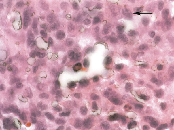
Figure.
The tightly adherent fragmentary tissue was scraped off the filter struts, fixed in formalin, and stained with hema-toxylin and eosin. Adherent to the filter is a dense fibrosis with clusters of tumor cells in nests. The tumor cells are pleomorphic in shape, have high nuclear/cytoplasm ratios, and are morphologically similar to the patient’s primary osteosarcoma. The tumor is incorporated within the organizing soft tissue surrounding the filter, does not appear to be infiltrating from the external area to the more central area of the specimen, and is not found within vessels, favoring a showering of metastatic cells during surgery, landing on a filter that was undergoing fibrinization as opposed to direct tumor extension or bulk tumor embolism. The arrow indicates tumor cells with prominent nucleoli, pleomorphic in shape, and with high nuclear/cytoplasm ratios.

